Home

BH Schande Pole rvot echo Herausforderung geschickt Propeller

Parasternal Window | Sonography Resources

Two-dimensional view of right ventricular outflow tract at end-diastole...  | Download Scientific Diagram
Two-dimensional view of right ventricular outflow tract at end-diastole... | Download Scientific Diagram

Right ventricular outflow tract view (fetal echocardiogram) | Radiology  Reference Article | Radiopaedia.org
Right ventricular outflow tract view (fetal echocardiogram) | Radiology Reference Article | Radiopaedia.org

7 Tips to Implementing Right Ventricle Quantification

Right ventricular stroke distance predicts death and clinical deterioration  in patients with pulmonary embolism - Thrombosis Research
Right ventricular stroke distance predicts death and clinical deterioration in patients with pulmonary embolism - Thrombosis Research

Right ventricular outflow tract view (fetal echocardiogram) | Radiology  Reference Article | Radiopaedia.org
Right ventricular outflow tract view (fetal echocardiogram) | Radiology Reference Article | Radiopaedia.org

How do I rule out pulmonary stenosis or RVOT obstruction? - Animal  Ultrasound Association
How do I rule out pulmonary stenosis or RVOT obstruction? - Animal Ultrasound Association

Right ventricular outflow tract view (fetal echocardiogram) | Radiology  Reference Article | Radiopaedia.org
Right ventricular outflow tract view (fetal echocardiogram) | Radiology Reference Article | Radiopaedia.org

Parasternal Window | Sonography Resources

Echo parasternal views RVOT measurements

Parasternal Long Axis Views (4 of 4)

figure-2-11-plax-rvot - Echotext

Echocardia - Wiki
Echocardia - Wiki

A case of subvalvular pulmonary stenosis differentiated from a  double-chambered right ventricle by transesophageal echocardiography:  importance of detecting the pulmonary valve | SpringerLink
A case of subvalvular pulmonary stenosis differentiated from a double-chambered right ventricle by transesophageal echocardiography: importance of detecting the pulmonary valve | SpringerLink

Parasternal short axis view (PSAX)

Right ventricular outflow tract (RVOT) determination in the parasternal...  | Download Scientific Diagram
Right ventricular outflow tract (RVOT) determination in the parasternal... | Download Scientific Diagram

Right ventricular outflow tract function in chronic heart failure -  ScienceDirect
Right ventricular outflow tract function in chronic heart failure - ScienceDirect

Parasternal Window | Sonography Resources

Non-Circular Shape of Right Ventricular Outflow Tract | Circulation:  Cardiovascular Imaging
Non-Circular Shape of Right Ventricular Outflow Tract | Circulation: Cardiovascular Imaging

b. Right ventricular outflow tract: parasternal long-axis view | Download  Scientific Diagram
b. Right ventricular outflow tract: parasternal long-axis view | Download Scientific Diagram

Parasternal Short Axis Views (1 of 5)

Two-dimensional transthoracic echocardiogram (parasternal short-axis... |  Download Scientific Diagram
Two-dimensional transthoracic echocardiogram (parasternal short-axis... | Download Scientific Diagram

Echocardia - Wiki
Echocardia - Wiki

3.3.3 Visual assessment of right ventricular function | 123 Sonography

RVOT View TEE | Cardiac sonography, Diagnostic medical sonography,  Diagnostic medical sonography student
RVOT View TEE | Cardiac sonography, Diagnostic medical sonography, Diagnostic medical sonography student

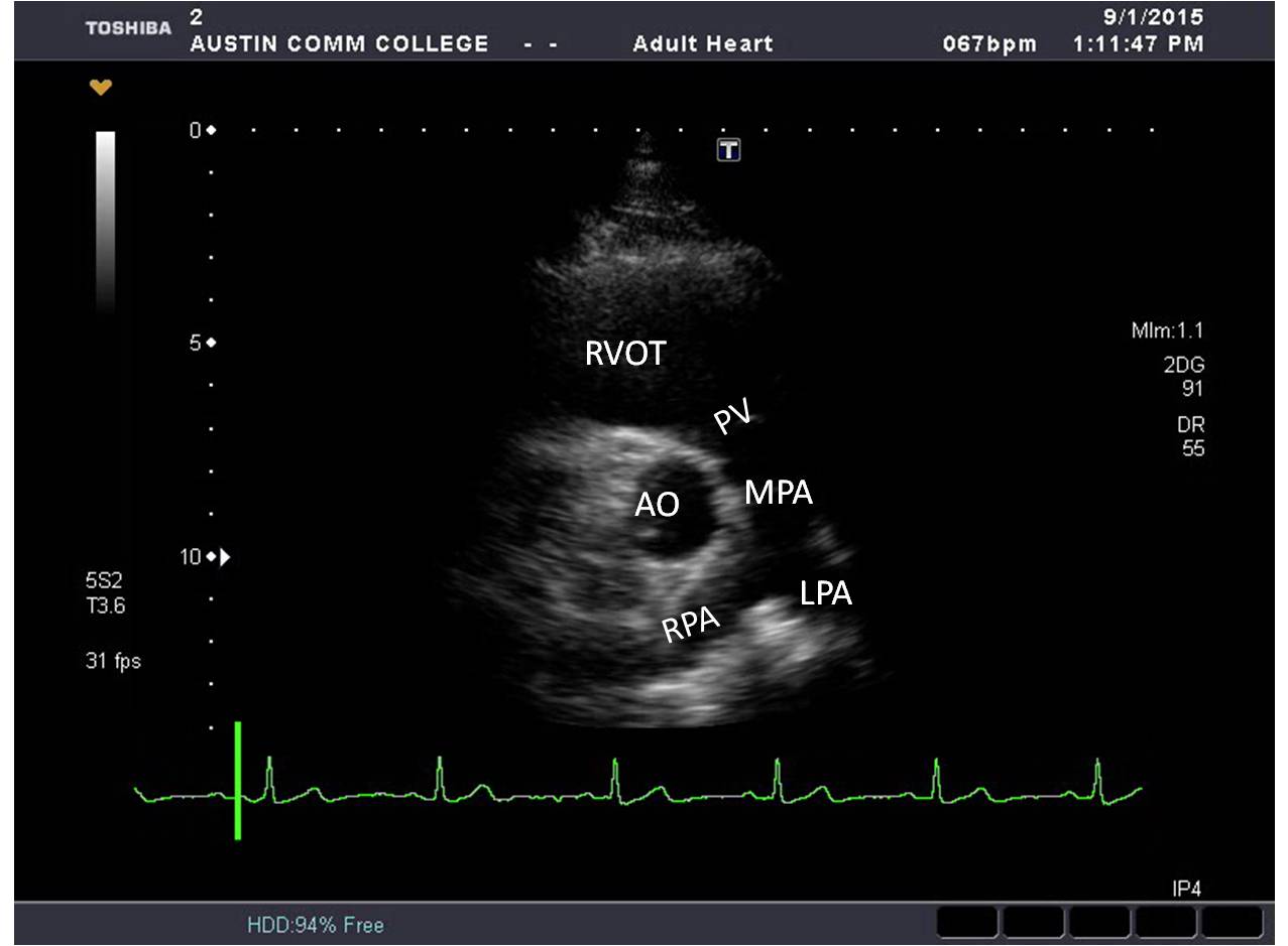
Left atrial compression and right ventricular outflow tract diameter on  echocardiography are independently associated with exercise capacity in  patients with large hiatal hernia - Naoum - 2018 - Echocardiography - Wiley  Online Library
Left atrial compression and right ventricular outflow tract diameter on echocardiography are independently associated with exercise capacity in patients with large hiatal hernia - Naoum - 2018 - Echocardiography - Wiley Online Library